
Blog
How to Reconstitute Research Peptides Correctly

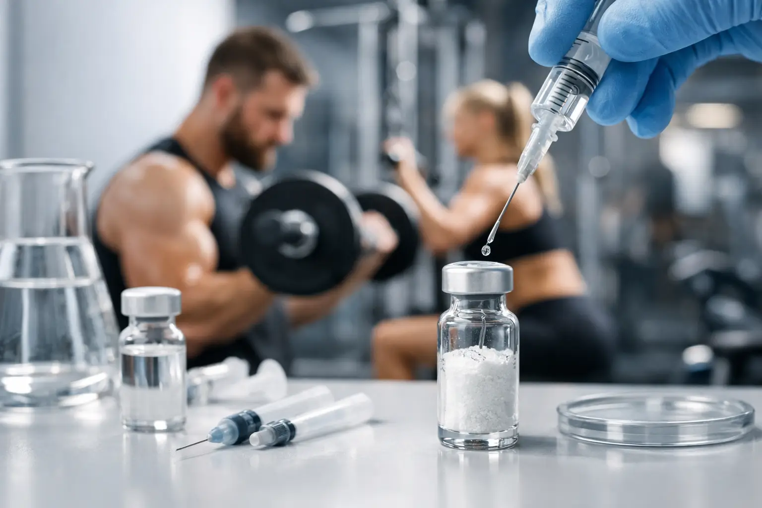
A lyophilized peptide vial is only as reliable as the preparation process that follows. Knowing how to reconstitute research peptides requires more than adding liquid to a vial. Solvent compatibility, final concentration, controlled handling, documentation, and storage conditions all affect whether a prepared research material remains suitable for consistent laboratory work.
Research peptides are supplied as experimental materials for laboratory use only. Preparation should be performed by qualified personnel using an established laboratory procedure, applicable safety controls, and the compound-specific documentation supplied with the material.
Start With the Vial Documentation
Before opening a vial, confirm the compound name, net peptide mass, lot identifier, storage instructions, and any solvent guidance provided for that material. Do not assume that one peptide’s reconstitution method applies to another. Molecular structure, solubility profile, formulation, and intended research concentration can vary substantially between products.
The vial should be inspected before preparation. A lyophilized peptide may appear as a powder, thin film, or compact cake at the base of the vial. Its appearance alone is not a final quality determination, but visible moisture, an unsealed closure, unusual discoloration, or damaged packaging should be documented and evaluated before use.
Keep the product label visible throughout the process. Once multiple vials and solvents are present on a work surface, identification errors become more likely than calculation errors. Controlled organization is a basic part of repeatable research handling.
Selecting a Compatible Reconstitution Solvent
The solvent is not an interchangeable accessory. Use the solvent identified in the applicable product documentation or research protocol. Sterile bacteriostatic water may be appropriate for certain materials, while other compounds may require a different diluent, such as acetic acid water, to support dissolution or stability under specified conditions.
Solvent selection depends on the peptide and the experimental requirements. A material that dissolves readily in one aqueous solvent may show incomplete dissolution, aggregation, or reduced stability in another. Do not select a solvent solely because it is commonly used with a different peptide.
Confirm that the solvent is within its stated shelf life and that its container has been stored according to its label. If working with a multi-use solvent container, maintain a record of the opening date and handling history. These details support traceability when results need to be reviewed later.
Calculate the Target Concentration Before Reconstitution
The amount of solvent added determines the final concentration of the prepared peptide solution. Establish that concentration before beginning, based on the requirements of the research protocol and the available vial mass.
The core calculation is straightforward:
Concentration = peptide mass divided by final solution volume
For example, reconstituting a vial containing 5 mg of peptide with 2 mL of compatible solvent produces a nominal concentration of 2.5 mg/mL. The same 5 mg vial reconstituted with 1 mL produces 5 mg/mL. The peptide mass remains unchanged; only the concentration changes.
Maintain unit consistency. A milligram is not interchangeable with a microgram, and a milliliter is not interchangeable with a microliter. Many preparation failures begin with a correct formula paired with mismatched units. Record the original vial mass, solvent volume, calculated concentration, date, and preparer’s initials in the laboratory record.
When a protocol requires multiple working concentrations, prepare them through documented dilution steps rather than relying on informal estimates. Each dilution should identify the source solution, dilution ratio, solvent used, final concentration, and storage conditions.
Prepare a Controlled Work Area
Reconstitution is best performed in a clean, organized environment using suitable laboratory practices. Gather the peptide vial, compatible solvent, sterile transfer equipment, labels, and documentation before beginning. Limiting interruptions reduces the chance of a wrong-vial, wrong-solvent, or wrong-volume event.
Allow refrigerated materials to equilibrate according to the applicable handling procedure before opening when condensation is a concern. Condensation around a stopper or cap can introduce avoidable uncertainty during transfer. Keep vial closures and transfer points protected from unnecessary contact.
Use appropriate personal protective equipment and aseptic technique consistent with the research setting. Avoid placing caps, stoppers, or sterile equipment on uncontrolled surfaces. If a vial stopper requires cleaning under the established procedure, allow the surface to dry fully before access.
How to Reconstitute Research Peptides With Minimal Stress
Using sterile transfer equipment, draw the calculated volume of compatible solvent. Introduce the solvent slowly down the inner wall of the peptide vial whenever practical. Direct, forceful delivery onto the lyophilized material can cause foaming or agitation, particularly with delicate peptide preparations.
After adding the solvent, allow the vial to stand briefly. Then mix gently by slow swirling or careful rolling between the fingers, if permitted by the material documentation. Do not shake the vial aggressively. Vigorous agitation may create foam, make visual assessment more difficult, and place unnecessary physical stress on the preparation.
Observe the solution under suitable lighting. A fully dissolved preparation should align with the expected appearance described in the relevant documentation. Persistent particles, cloudiness, unusual color, or visible fibers warrant a pause in the workflow. Do not attempt to force dissolution through repeated shaking, warming, or unverified solvent additions. Record the observation and follow the established quality procedure for the material.
Do not combine two research materials in a single vial unless the experimental protocol specifically calls for that preparation and compatibility has been established. A blend supplied as a formulated product is not equivalent to an improvised combination of separate compounds.
Label, Store, and Track the Prepared Solution
The reconstituted vial should be labeled immediately. At minimum, the label should identify the compound, lot number, solvent, final concentration, reconstitution date, and any protocol-specific storage condition. If more than one researcher has access to the material, add preparer identification and a beyond-use date defined by the applicable procedure.
Store the solution according to compound-specific instructions. Temperature, light exposure, freeze-thaw cycles, and container handling can affect stability. A refrigerator is not a universal answer, and repeated removal from controlled storage can introduce temperature variation that is easy to overlook.
Avoid keeping prepared solutions longer than the established research procedure allows. When stability data are unavailable or uncertain, a conservative handling approach and complete records are more defensible than assumption. Body Tech Peptides emphasizes controlled handling because material consistency depends on what occurs after a vial leaves its original packaging as much as on the original product standard.
Common Reconstitution Errors That Undermine Research Consistency
Several errors recur across peptide preparation workflows. The most common are using an unverified solvent, calculating concentration after rather than before solvent addition, misreading mass units, shaking the vial, and failing to label the prepared solution immediately.
Another frequent issue is relying on memory. A vial may look familiar while containing a different peptide mass or lot. Written records prevent an apparently small handling difference from becoming an unexplained variable in later work.
Temperature shortcuts are also risky. Do not apply heat, freeze a preparation, or alter storage conditions simply to accelerate dissolution unless those steps are expressly supported by the material documentation. Speed is not a substitute for controlled technique.
A well-prepared vial does not begin with the solvent transfer. It begins with verified documentation, a calculated concentration, a compatible diluent, and a record that another qualified researcher can follow without guesswork.

3 thoughts on “How to Reconstitute Research Peptides Correctly”